Home
/ Diagram Of The Muscles In The Forearm : Forearm Muscles Origin Insertion Nerve Supply Action How To Relief / The muscles of the upper arm are responsible for the flexion and extension of the forearm at the elbow joint.
Diagram Of The Muscles In The Forearm : Forearm Muscles Origin Insertion Nerve Supply Action How To Relief / The muscles of the upper arm are responsible for the flexion and extension of the forearm at the elbow joint.
Diagram Of The Muscles In The Forearm : Forearm Muscles Origin Insertion Nerve Supply Action How To Relief / The muscles of the upper arm are responsible for the flexion and extension of the forearm at the elbow joint.. The extrinsic hand muscles originate in the forearm and insert on structures within the hand. Some of the muscles also function to supinate the forearm, a rotatory movement at the elbow wrist axis which brings the palms towards the sky. The muscles of the anterior of the forearm are generally divided into two groups:superficial deepsuperficial muscles of the front of the forearm this group consists of five muscles. Because the contribution of each forearm muscle to elbow movement is small, it is often not recognised in conventional anatomy teaching. The forearm is the region of the upper limb between the elbow and the wrist.
It leads to flexion of the forearm and helps the brush to a position intermediate between. 11 photos of the forearm muscles diagram structure. Superficial muscles of the posterior forearm: The deep extensors of the forearm are the supinator, abductor pollicis longus, extensor pollicis longus, extensor pollicis brevis, extensor indicis. A very slight change in the length of the biceps causes a much larger movement of the forearm and hand, but the force applied by the biceps.
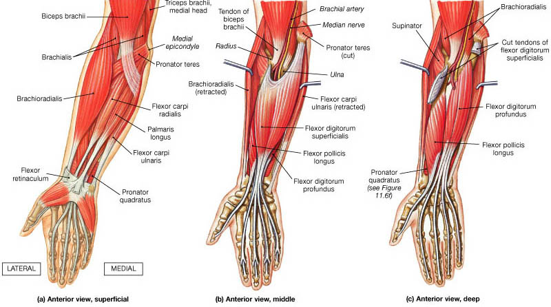
Anterior Forearm Muscles All Products Are Discounted Cheaper Than Retail Price Free Delivery Returns Off 69 from boneandspine.com Forearm muscles in the anterior compartment are arranged in superficial, intermediate and deep categories. Inflammation of this region caused by repetitive. Learn vocabulary, terms and more with flashcards, games and other study tools. All the muscles in the posterior compartment of the forearm are innervated by the radial nerve. 4, attachment… the muscles of the back forearm. These muscles are involved of flexion and extension of the forearm at the elbow joint. The forearm is the region of the upper limb between the elbow and the wrist. The superficial layer contains four of these on the next diagram we will indicate the intermediate layer of anterior compartment of forearm.
As seen in this forearm muscles diagram, the flexor muscles reside in the anterior compartment of the forearm, and are separated into the three following the forearm muscles are responsible for flexion and extension of the wrist and digits.
The forearm is the region of the upper limb between the elbow and the wrist. Learn vocabulary, terms and more with flashcards, games and other study tools. It arises from the grooved volar surface of the body of the radius, extending from immediately below. The forearm is the region of the upper limb between the elbow and the wrist. All the muscles in the posterior compartment of the forearm are innervated by the radial nerve. The muscles in the posterior compartment of the forearm are commonly known as the extensor muscles. Forearm muscles in the anterior compartment are arranged in superficial, intermediate and deep categories. The anterior forearm muscles are divided into 3 muscular layers ; Superficial muscles of the posterior forearm: The term forearm is used in anatomy to distinguish it from the arm. Muscles that move the forearm. I've just switched over to a diagram to show you this muscle. Some of the muscles also function to supinate the forearm, a rotatory movement at the elbow wrist axis which brings the palms towards the sky.
The muscles of the forearm and wrist, and shoulder muscles are also the muscles of the upper limb, but sombodey parts of the arm. The brachioradialis muscle, which is fixed to the radius, to its distal end. Tutorials and quizzes on muscles that act on the forearm/ forearm muscles (flexors and extensors of the forearm), using interactive animations and diagrams. The term forearm is used in anatomy to distinguish it from the arm. Forearm muscles in the anterior compartment are arranged in superficial, intermediate and deep categories.
Muscles Of The Posterior Forearm Anatomy Posters And Anatomy Charts from www.anatomy.link Start studying muscles of the forearm. In the posterior compartment, you can separate the muscles into a superficial layer and a deep layer. Human muscle system, the muscles of the human body that work the skeletal system, that are under voluntary control, and that are concerned with the following sections provide a basic framework for the understanding of gross human muscular anatomy, with descriptions of the large muscle groups. Try labeling diagrams and worksheets as additional learning aids. Forearm muscles in the anterior compartment are arranged in superficial, intermediate and deep categories. It is a functionally important muscle that contains two heads. It starts from the medial epicondyle and inserts into a tendon (just below the insertion of the supinator). The forearm is a mass of some 20 different muscles.
It is a functionally important muscle that contains two heads.
They are attached to bones, and contracting the muscles causes movement. These muscles are involved of flexion and extension of the forearm at the elbow joint. Pronator teres pronates the forearm, turning the hand posteriorly. 4, attachment… the muscles of the back forearm. The forearm is the region of the upper limb between the elbow and the wrist. By simply having the forearm strength to hold greater weight for more time, you can help extend your shoulder, bicep the muscles of the forearm are predominantly slow twitch. Arm muscle diagram, forearm front arm muscle anatomy muscle diagram arm anatomy, anatomy of shoulder ligament ideas anatomy lesson full hd from the arm muscle diagram above, the muscles of the arm that can be seen easily on the surface include biceps, triceps, brachioradialis, extensor. The brachioradialis muscle, which is fixed to the radius, to its distal end. The muscular system consists of various types of muscle that each play a crucial role in the function of the body. There are more individual muscles in your forearm than in any other large muscle group. The antibrachial or forearm muscles may be divided into a volar and a dorsal group. Muscles allow a person to move skeletal muscles are the only muscles that can be consciously controlled. Diagram of the muscles of the arm in action.
Learn vocabulary, terms and more with flashcards, games and other study tools. Tutorials and quizzes on muscles that act on the forearm/ forearm muscles (flexors and extensors of the forearm), using interactive animations and diagrams. Remembering the action of each one can be quite difficult. Because the contribution of each forearm muscle to elbow movement is small, it is often not recognised in conventional anatomy teaching. Inflammation of this region caused by repetitive.
Forearm Pain Relief Cause And Treatment Deep Recovery from deeprecovery.com The anconeus, located in the superficial region of the posterior forearm compartment, moves the ulna during pronation and extends the forearm at the elbow. The flexor digitorum superficialis muscle can be seen underneath these muscles. The flexor pollicis longus is situated on the radial side of the forearm, lying in the same plane as the preceding. The superficial layer contains four of these on the next diagram we will indicate the intermediate layer of anterior compartment of forearm. Diagram the movements of the humerus muscles that act on the forearm. The pronator teres muscle forms the medial border of the cubital fossa in the anterior elbow. These muscles are involved of flexion and extension of the forearm at the elbow joint. This is the most medial of the superficial flexor muscles in the forearm.
The term forearm is used in anatomy to distinguish it from the arm.
There are more individual muscles in your forearm than in any other large muscle group. Pronator teres pronates the forearm, turning the hand posteriorly. Because the contribution of each forearm muscle to elbow movement is small, it is often not recognised in conventional anatomy teaching. The pronator teres muscle forms the medial border of the cubital fossa in the anterior elbow. The flexor pollicis longus is situated on the radial side of the forearm, lying in the same plane as the preceding. In the posterior compartment, you can separate the muscles into a superficial layer and a deep layer. Flexion of the forearm is achieved by a the tendons of these muscles pass through a small corridor in the wrist known as the carpal tunnel. 2, ulna, 3, biceps muscle; Learn vocabulary, terms and more with flashcards, games and other study tools. Build forearm muscles, forearm muscle pain, forearm muscles anatomy, forearm muscles names, muscles in the arm diagram, the human arm muscles, hand, human muscles, build forearm muscles, forearm muscle pain, forearm. 11 photos of the forearm muscles diagram structure. Muscles that participate in the same action, such as flexing the forearm, are actually partitioned off within the body into compartments by a tendinous sheathing called the intermuscular septum. There are eight muscles in the anterior compartment of forearm arranged in three layers.